Please click the button below to go to our email login page
|
Kidney Organoids Secure 14.3-Point Article! Zhihong Liu’s Team from Nanjing University Reveals New Directions for Kidney Disease TreatmentOrganoids, derived from the body's own tissues or stem cells, are 3D structural model with original tissues and organs formed via in vitro 3D culture.
Today, we are going to share a paper published on Advanced Science with IF of 14.3, hoping to inspire you from different perspectives.
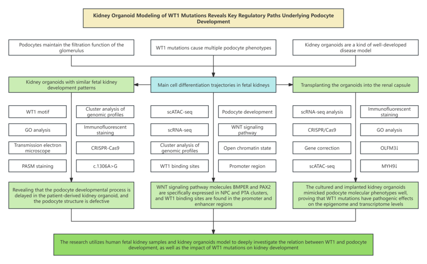
1. Research background 1.1 Podocytes are a special cell type in the kidney and are responsible for maintaining the filtration function of the glomerulus. 1.2 During the development of kidney, the renal cell carcinoma suppressor gene (WT1) expression is enhanced along with the development of metanephric mesenchyme and podocyte progenitors, and ultimately limited to mature podocytes. 1.3 WT1 mutations result in multiple podocyte phenotypes accompanied with defect of glomerular basement membrane. 1.4 Kidney organoids are a kind of well-developed disease model for exploring the etiology of gene mutations in patients.
2. Technical routes
3. Research results 3.1 Identifying WT1‐related epigenomic landscape of human podocyte development in main cell differentiation trajectories in fetal kidney. 3.2 Identifying WT1‐related epigenomic landscape of human podocyte development in main cell differentiation trajectories in cultured kidney organoids. 3.3 Dysregulated cellular processes in the SSBPod and podocytes of cultured kidney organoids induced by WT1 mutations 3.4 WT1 mutations-induced dysregulated cellular processes in podocytes of implanted kidney organoids. 3.5 The podocyte phenotype was rescued by CRISPR/Cas9 editing of patient-derived kidney organoids carrying the WT1 mutation.
4. Conclusion This research generates single-cell chromatin accessibility and gene expression maps of fetal kidneys and kidney organoids. WT1‐targeted genes play a vital role in maintaining podocyte development and structure. To further clarify the functional significance of WT1-mediated transcriptional regulation, they culture and implant patient‐derived kidney organoids derived from patient‐iPSCs that carried a heterozygous missense mutation in WT1. scRNA-seq and functional analysis results demonstrated that WT1 mutations cannot activate targeting genes MAGI2, MYH9 and NPHS1, resulting in delayed development of podocyte and damaged cell structure. Notably, CRISPR-Cas9 gene editing can be applied to correct mutations in iPSCs and thus rescue podocyte phenotype. Collectively, this research illuminated the epigenomic landscape of WT1 related to human podocyte development, and identified the pathogenic effect of WT1 mutations This research not only provides new insights into kidney development mechanism, but also offer new directions to kidney disease study and treatment. |


